| book contents |
chapter contents | |
| → |
|
♦ Reproduction - Testis; Normal Histology - testis development
|
|
|
|
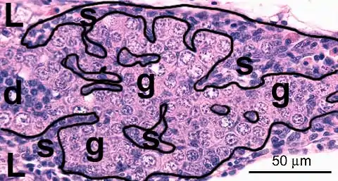
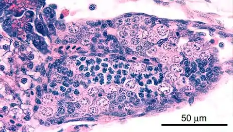
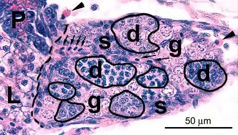
2. early development
An early feature of male differentiation of the gonad is ingrowth of stroma [s]. Other features, which are not present on this image are appearance of a clustered organisation (spermatocysts) and lumen formation. This gonad mainly contains undifferentiated gonocytes [g], but early stages of differentiation [d] are also present. A small part of the liver [L] is visible. |
|
|
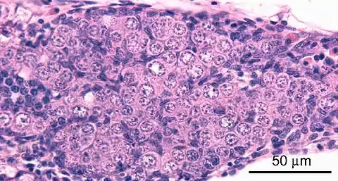
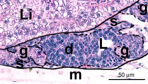
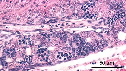
3. early development
Adjacent structures in this image are: liver [Li] on the medial side mesenchyme [m] on the lateral side
|
|
|
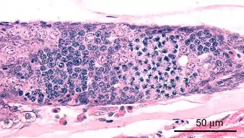
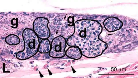
4. early development
Early features of male differentiation of the gonad are the appearance of a clustered organisation (spermatocysts) and ingrowth of stroma [s]. This gonad contains early stages of differentiation [d], as well as undifferentiated gonocytes [g]. A vessel [arrows] can be observed, and adjacent structures in this image are: liver [L], pancreas [p], and peritoneal cells [arrowhead] |
|
|
5. early development
This gonad shows the combined presence of clustered organisation (spermatocysts [d]) and lumen formation [arrows], as indications of early male differentiation. Early stages of differentiation are predominant [d], but undifferentiated gonocytes [g] are also abundant. Adjacent structures in this image are: liver [L] peritoneal cells [arrowheads] |
|
|
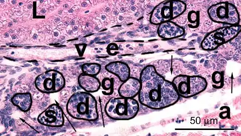
6. intermediate development
This gonad shows the combined presence of clustered organisation (spermatocysts [d]) and lumen formation [arrows] as indications of early male differentiation. A prominent blood vessel [v] filled with erythrocytes [e] is present in the stroma. The spematogenic epithelium contains mainly early and intermediate stages of differentiation [d], up to early spermatids [s]; clusters of undifferentiated gonocytes [g] are also present. Adjacent structures in this image are: liver [L] abdominal wall [a] |
|
|
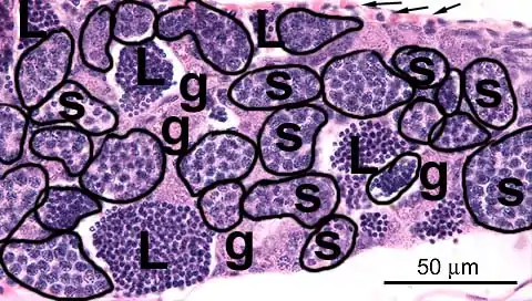
7. advanced development
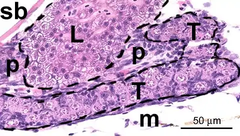
This gonad can be identified as a testis by the presence of well-developed lumina, which are filled with sperm [L], and by the clustered organisation (spermatocysts [s]). Clusters of spermatogonia [g] are also present. Adjacent structures in this image are: clustered peritoneal cells [arrows] |
|
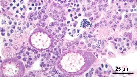
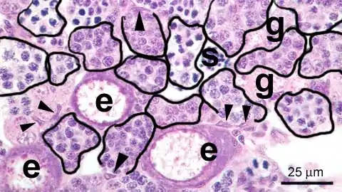
♦ testis-ovaEarly oocytes [e] are observed in the developing testis at varying incidences (1:350 in the RIVM zebrafish stock, 1:10 in the stock of the Veterinary Faculty of the Utrecht University). Thise image shows the phenomenon at a relatively more advanced developmental stages, which includes mature sperm [s]. There is an obvious clustered organisation (spermatocysts), combined with clusters of spermatogonia [g]. Incidently, Sertoli cells [arrowheads] can be discerned. The presence of primitive oocytes in the developing testis has been interpreted as a feature of undifferentiated gonochorism or protogyny: the undifferentiated gonad develops into an ovary-like gonad in all individuals and then in about one-half of the population transforms into a testis (Takahashi-H. Juvenile hermaphroditism in the zebrafish, Brachydanio rerio. Bull.Fac.Fish.Hokkaido Univ.28:57-65;1977). Takahashi describes "intersexuality" similar to the images presented here with a maximal incidence of 1:4. These varying incidences indicate that the balance of sex-determining factors is delicate and environmental or genetical variations between stocks may produce a slightly different differentiation process; furthermore. Our observations do not suppport the undifferentiated gonochoristic or protogyny paradigm in zebrafish. |
|

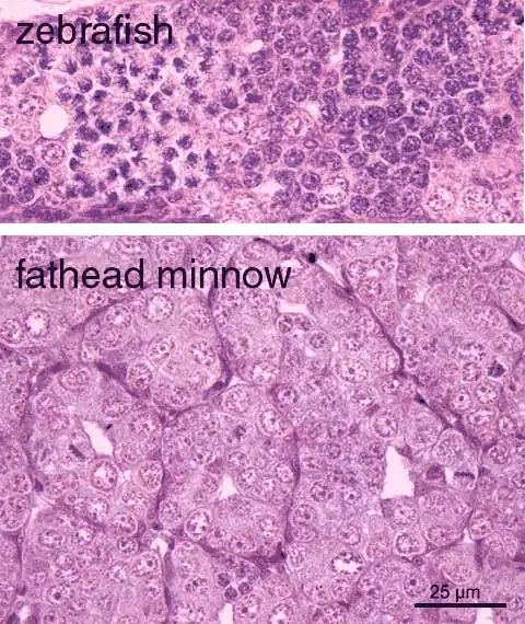
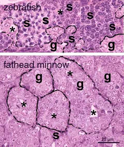
♦ variability between species
| |
|
|
|
|
age of the zebrafish is appr. 6w and of the fathead minnow appr. 9w; both images have equal magnification |
|
|
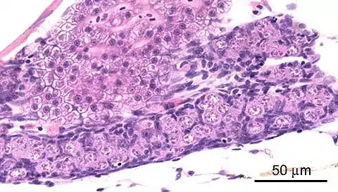
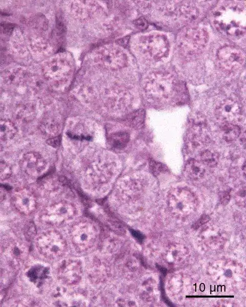
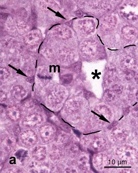
detail of early fathead minnow testis | |
|
|
|
|
age of the fathead minnow appr. 9w; fathead minnow sections were kindly provided by Dr. Grace Panter |
|
|
♦ development of testis orientation in the peritoneal cavity
| ||
|
|
|
|


|
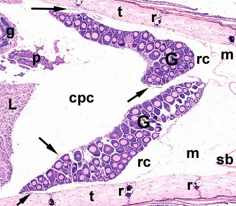
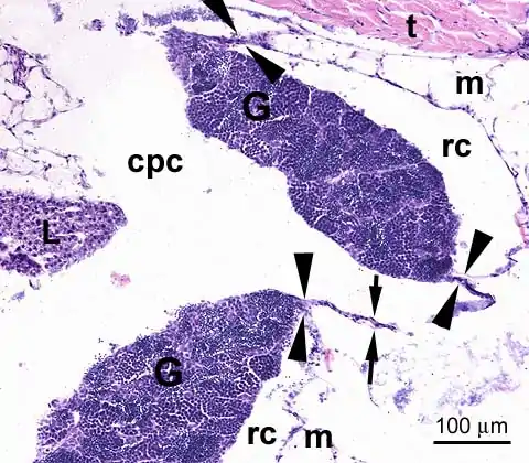
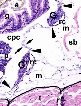
persisting bilateral attachment in the ovary [Fig.A4]: In ovary development, the retrogonadal cavity [rc] persists and even expands; eventually it becomes the oviduct. The ovary [G] thus remains attached bilaterally to the peritoneal wall. The peritoneum [arrows] constitues the boundary between this future oviduct and the peritoneal cavity [cpc]. |
|
|
|
|
|
Other structures in these image are: liver [L], swim bladder [sb], gut [g] (note Artemia salina eggshell remains [a] in the gut lumen in Fig.A2), pancreas [p], trunk wall musculature [t], which shows its compartimental organization in somites [outlined], and which contains a rib [r] between two somites, retroperitoneal mesenchyme [m]. |
|